
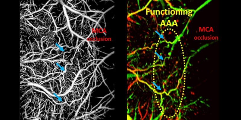
Image: Monitoring arteriogenesis at the cerebral arteriolo-arteriolar anastomoses (AAAs) in mouse brain during middle cerebral artery occlusion using OCT-based microangiography (OMAG) (left) and Doppler OMAG (right).
Abstract
Background
Arteriogenesis describes the active growth of the pre-existing collateral arterioles, which is a crucial tissue-saving process in occlusive vascular diseases.
New method
We propose to use optical coherence tomography (OCT)-based microangiography (OMAG) to monitor arteriogenesis following artery transection in mouse ear and focal stroke in mouse brain.
Results
Our longitudinal mouse ear study shows that the growth phase of arteriogenesis, indicated by changes in collateral vessel diameter and velocity, occurs between 12 and 24 h after vessel transection. Additionally, the magnitude of local inflammation is consistent with the time course of arteriogenesis, judging by the tissue thickness measurement and lymphatic vessel signals in OCT. In the mouse brain study, collateral vessel morphology, blood flow velocity and directionality are identified, and an activation of the collateral flow at the arteriolo-arteriolar anastomoses (AAA) is observed during stroke.
Comparison with existing methods
In comparison with histology and fluorescence imaging, OCT/OMAG is completely non-invasive and capable of producing consistent results of longitudinal changes in collateral vessel morphology and vasodynamics.
Conclusion
OCT/OMAG is a promising imaging tool for longitudinal study of collateral vessel remodeling in small animals. This technique can be applied in guiding the in vivo experiments of arteriogenesis stimulation to treat occlusive vascular diseases, including stroke.