
$2.9M NIH grant will support advances in 3D printing of functional human liver tissue and $1M Keck grant funds new approaches to look at how disease scarring disrupts liver regeneration.
NIH: 3D printing human liver tissue
Kelly Stevens, assistant professor of bioengineering and of laboratory medicine and pathology, has received a $2.9 million grant from the National Institutes of Health for a four-year project aimed at 3D printing functional human liver tissue.
“This support will bring us closer to our dream of 3D printing human livers for clinical transplantation,” said Dr. Stevens, who is also a researcher at the UW Medicine Institute for Stem Cell and Regenerative Medicine (ISCRM)
The 3D printed tissue could also serve as an interim treatment or alternative for people who need liver transplantation.
In 2019, Dr. Stevens and her collaborator, Jordan Miller, an associate professor of bioengineering at Rice University, created a breakthrough technique for bioprinting 3D tissues with exquisitely entangled vascular networks that mimic the body’s natural passageways for blood, air, lymph and other vital fluids. They later created new formations of light-absorbing “bioinks” that increased the resolution of 3D-printed vasculature. They also found they could increase the density of cells in 3D-printed tissues by adding biological matrices to the bioinks.
The Stevens lab and collaborators will work to scale up their system, printing larger tissues with a greater density of blood vessels to keep the densely packed liver cells alive. They will work to formulate a new library of bioinks and show that their printed tissues can carry out liver function. By the end of the project, they hope to 3D print human liver tissue and show that it can function well enough to rescue mice with liver injuries.
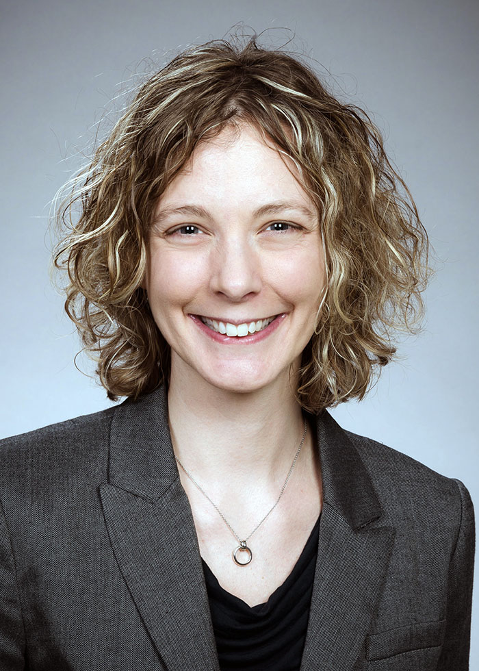
UW bioengineer and regenerative medicine researcher Kelly Stevens
Keck: Mechanical properties of liver regeneration
Dr. Stevens has also won a $1 million grant from the W.M. Keck Foundation to advance her team’s work to understand the factors that interfere with liver regeneration.
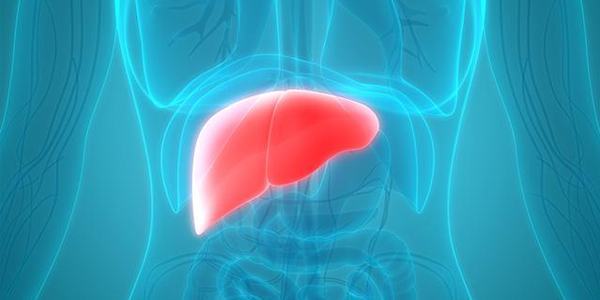
The liver is one of the most complex organs in the body and performs a multitude of vital roles. The liver is also the only visceral organ that can regrow itself – at least when healthy.
Unlike healthy livers, those damaged by disorders like cirrhosis have difficulty repairing themselves. Why scarring from disease or toxins diminishes the remarkable ability of a living liver to regenerate is not yet understood.
Most efforts to explore liver regeneration – or its failure – have looked at biochemical and genetic factors, Dr. Stevens explained, because scientific tools for these studies are easily available.
Instead, Dr. Stevens will take on the challenge of developing new methods to examine other factors. She wants to examine the fundamental role of mechanical forces in shaping or hindering the re-formation of livers.
“Mechanobiology has been a major missing piece in liver regeneration research,” she said.
Increasingly, Dr. Stevens pointed out, scientists are finding, in other areas of study, that the mechanical properties of the environment where cells live can have profound effects on their behaviors, motility, lineage pathways, proliferation and functions.
Dr. Stevens proposed that the stiffness of a cirrhotic liver might have a mechanical influence on hepatic cell destinies.
“When chronically injured livers become cirrhotic and stiffened, liver regeneration fails,” she noted, “yet cirrhotic or stiffened livers frequently progress to cancer.” This paradox suggests stiffer environments may stimulate proliferation of cells in some situations.
Testing hundreds of microenvironments at once
The Stevens lab is taking a new angle to a so far intractable challenge. Her lab has developed a model of human liver regeneration. In it, artificial liver seed, produced by 3D printing, are grafted into mice who have liver disease. These seeds expand and enable the creation of designer human liver tissues. Varying degrees of mechanical stiffness can be tested.
These models become part of a system to investigate the effects of mechanical cues on human liver cells representing different disease states. The cell populations and their surrounding environment can be disrupted to evaluate the effects on growth.
A drawback of the original testing system is that only one tissue could be implanted. The Stevens team will apply a newer method, highly parallel tissue grafting (HPTG), to allow the scientists to simultaneously probe the effects of hundreds of different mechanical microenvironments on human liver regeneration.
Human liver cell biobank
Dr. Stevens and her team hypothesize that liver cirrhosis prohibits regeneration either because the physical constraints of the stiffened liver don’t allow for liver cells to expand their numbers, or because the liver cells have somehow reached their limits to proliferate. The researchers plan to establish a biobank of human liver cell samples representing a spectrum of disease states, and a portfolio of materials representing a range of mechanical stiffness, from normal liver to cirrhosis.
With collaborative consultations from UW Medicine experts in other fields, the project will incorporate high dimensional statistical analysis, computational biology, and machine learning advances to model spatial variation in liver regeneration.
The researchers hope their latest studies will demystify the mechanisms of liver regeneration. In addition to identifying how environmental mechanical factors interact with cellular disease to interfere with liver regeneration, they also would like to discover other useful findings. For example, what they learn might advance the engineering of artificial tissues as a potential therapy for liver failure. It perhaps might lead as well to ways to boost the regenerative potential of the damaged liver itself.
The scientists would like to see the technological innovations from this study have wider uses in biomedical science. These might include such areas as heart regenerative research or studies of cancer growth.
The W. M. Keck Foundation was established in 1954 in Los Angeles by William Myron Keck, founder of The Superior Oil Company. One of the nation’s largest philanthropic organizations, the W. M. Keck Foundation supports outstanding science, engineering and medical research. The Foundation also supports undergraduate education and maintains a program within Southern California to support arts and culture, education, health and community service projects.
Keck award adapted from a UW Medicine release by Leila Gray.
Read more about Kelly Stevens’ Keck award and her research in this ISCRM article.