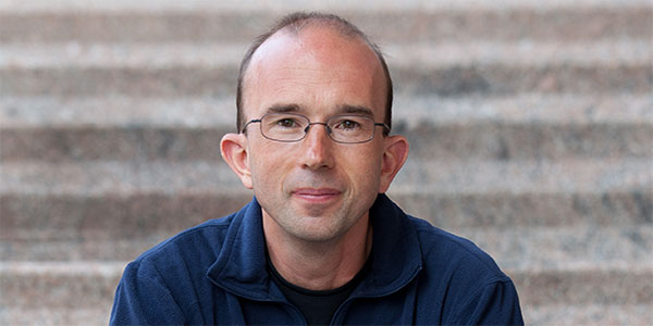
Joint Professor of Pediatrics and Bioengineering
studholm@uw.edu
Phone: (206) 221-7022
Office: HSB RR439A
Colin Studholme
Methods
MR image acquisition and reconstruction during subject motion,
Image resolution enhancement in structural and diffusion imaging
Algorithms for automated tissue segmentation and anatomical parcellation
Biomedical shape and pattern analysis,
Graph methods for studying network connectivity and its change over time.
Spatial statistics for brain mapping
Applications
Early human brain tissue growth patterns
The development of fetal brain functional and structural connectivity
The impact of genes and the environment on early brain development
Imaging biomarkers to improve the management of pregnancies and premature birth
Msc, Edinburgh University, Scotland, 1991
BEng, Bradford University, England, 1990
BIOEN 299
M. Fogtmann, S. Seshamani, C. Kroenke, X. Cheng, T. Chapman, J. Wilm, F. Rousseau, M. Koob, J.-L. Dietemann, C. Studholme, “A unified approach to diffusion direction sensitive slice registration and 3D DTI reconstruction from moving fetal brain anatomy”, IEEE Trans. Med. Imaging, Vol. 33, No. 2, February 2014
C. Studholme, F. Rousseau, “Quantifying and modelling tissue maturation in the living human fetal brain”, International Journal of Developmental Neuroscience, Volume 32, February 2014, Pages 3-10, ISSN 0736-5748
J. Scott, V. Rajagopalan, P.A. Habas, K. Kim, A.J. Barkovich, O.A. Glenn, C. Studholme, ” Volumetric and surface-based 3D MRI analyses of fetal isolated mild ventriculomegaly Brain Struct Funct, 2012
V. Rajagopalan, J. Scott, P.A. Habas, K. Kim, F. Rousseau, A.J. Barkovich, O.A. Glenn, C. Studholme, “Mapping directionality specific volume changes using tensor based morphometry: An application to the study of gryrogenesis and lateralization of the human fetal brain,” NeuroImage, Volume 63, Issue 2, 1 November 2012, Pages 947-958
P. A. Habas, J. A. Scott, A. Roosta, V. Rajagopalan, K. Kim, F. Rousseau, A. J. Barkovich, O. A. Glenn, and C. Studholme, “Early folding patterns and asymmetries of the normal human brain detected from in utero MRI,” Cereb. Cortex, vol. 22, no. 1, pp. 13-25, 2012.
C. Studholme, “Mapping fetal brain development in utero using MRI: the big bang of brain mapping,” Annu. Rev. Biomed. Eng., vol. 13, no. 1, pp. 345-368, August 2011.
V. Rajagopalan, J. Scott, P.A. Habas, K. Kim, F. Rousseau, A.J. Barkovich, O.A. Glenn, C. Studholme, “Mapping directionality specific volume changes using tensor based morphometry: An application to the study of gryrogenesis and lateralization of the human fetal brain,” NeuroImage, Volume 63, Issue 2, 1 November 2012, Pages 947-958.
P. A. Habas, J. A. Scott, A. Roosta, V. Rajagopalan, K. Kim, F. Rousseau, A. J. Barkovich, O. A. Glenn, and C. Studholme, “Early folding patterns and asymmetries of the normal human brain detected from in utero MRI,”, Cereb. Cortex, vol. 22, no. 1, pp. 13-25, 2012.
C. Studholme, “Mapping fetal brain development in utero using MRI: the big bang of brain mapping,” Annu. Rev. Biomed. Eng., vol. 13, no. 1, pp. 345-368, August 2011.
V. Rajagopalan, J. A. Scott, P. A. Habas, J. M. Corbett-Detig, K. Kim, F. Rousseau, A. J. Barkovich, O. A. Glenn, and C. Studholme, “Local tissue growth patterns underlying normal fetal human brain gyrification quantified in utero,” J. Neurosci., vol. 31, no. 8, pp. 2878-2887, February 2011.
P. A. Habas, K. Kim, J. M. Corbett-Detig, F. Rousseau, O. A. Glenn, A. J. Barkovich, and C. Studholme, A spatiotemporal atlas of MR intensity, tissue probability and shape of the fetal brain with application to segmentation,” Neuroimage, vol. 53, no. 2, pp. 460-470, November 2010.
K. Kim, P. A. Habas, F. Rousseau, O. A. Glenn, A. J. Barkovich, and C. Studholme, “Intersection based motion correction of multislice MRI for 3-D in utero fetal brain image formation,” IEEE Trans. Med. Imaging, vol. 29, no. 1, pp. 146-158, January 2010.
C. Studholme, “Dense Feature Deformation Morphometry: Incorporating DTI Data into Conventional MRI Morphometry”, Medical Image Analysis, Volume 12, Issue 6, December 2008, Pages 742-751 2008.
F. Rousseau, O. Glenn, B. Iordanova, Cl. Rodriguez, D. Vigneron, J. Barkovich, C. Studholme, Registration Based Approach for Reconstruction of High Resolution In Utero MR Brain Images, Academic Radiology, 13(9), Sept 2006, pp 1072-1081.
C. Drapaca, V. Cardenas, C. Studholme, “Segmentation of Tissue Boundary Evolution from Brain MR Image Sequences using Multi-phase Level Sets”, Computer Vision and Image Understanding, volume 100, 2005, pages 312-329.
C.Studholme, V. Cardenas, “A Template Free Approach to Volumetric Spatial Normalisation of Brain Anatomy”, Pattern Recognition Letters, Vol 25(10), Jul 2004, pp. 1191-1202.
C.Studholme, V. Cardenas, R. Blumenfeld, N. Schuff, H.J. Rosen, B. Miller, M. Weiner, “A Deformation Tensor Morphometry Study of Semantic Dementia with Quantitative Validation”, NeuroImage, Vol 21, pp 1387-1398, April 2004.
C. Studholme, E. Novotny, I.G. Zubal, J.S. Duncan, Estimating Tissue Deformation Between Functional Images Induced by Intracranial Electrode Implantation Using Anatomical MRI, NeuroImage, Vol 13(4), pp 561-576, 2001.
C. Studholme, T.C. Constable, J.S. Duncan, Non-Rigid Spin Echo MRI Registration Incorporating an Image Distortion Model: Application to Accurate Alignment of fMRI to Conventional MRI, IEEE Trans. Med. Imaging, Vol 19, No 11. Nov 2000.
C. Studholme, D.L.G.Hill, D.J. Hawkes, An Overlap Invariant Entropy Measure of 3D Medical Image Alignment, Pattern Recognition, Vol. 32(1), Jan 1999, pp 71-86.
C.Studholme, D.L.G.Hill, D.J. Hawkes, Automated 3-D Registration of MR and CT Images of the Head, Medical Image Analysis, Vol. 1(2)., 1996, pp 163-175.
D.L.G. Hill, D.J. Hawkes, T.C.S. Cox, A.J. Strong, W.L. Wong, C.F. Ruff, N. Kitchen, D.G.T. Thomas, J.E. Cross-man, C. Studholme, A.J. Gandhe, S.E.M. Green, G.P. Robinson, Accurate Frameless Registration of MR and CT Images of the Head: Applications in Surgery and Radiotherapy Planning, Radiology, 191, 1994, pp 447-454.
In the News
New technology images fetal brain activity in 4D
2022-07-13T14:01:14-07:00March 13th, 2017|
Colin Studholme to present 2015-16 UW Medicine Science in Medicine Lecture
2021-01-21T06:04:32-08:00February 10th, 2016|


