


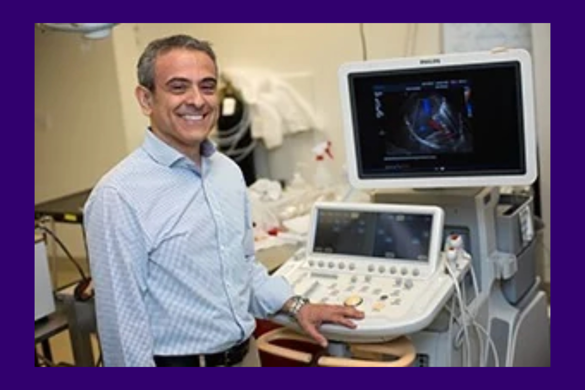
 UW Bioengineering Professor Mike Averkiou and his collaborators received a grant from the Andy Hill CARE Fund to develop a new approach to detecting liver cancer – and are already moving it toward the marketplace.
Current methods of diagnosing liver cancer come with “lots of maybes,” according to Professor Mike Averkiou. And those “maybes” about the existence, type, and size of a tumor come at a price. A recent study in the journal Clinical Gastroenterology and Hepatology showed that late detection of liver cancer significantly limits treatment options for patients.
“Liver cancer is hard to detect, and that makes it hard to treat,” whether with surgery or with minimally invasive approaches such as microwave, radio, or cryo-ablation. Doctors also have difficulty determining the margins where a tumor and healthy tissue meet and identifying smaller, nearby “satellite” tumors. A widely accepted technique called LI-RADS (Liver Imaging Reporting and Data System) detects tumors based on their blood flow patterns. While LI-RADS delivers relatively few false-positives, it fails to detect tumors early in their progression.
“This is a big problem,” Averkiou said.
Averkiou and his colleagues in the UW School of Medicine recently began work on a new way of imaging liver cancer. It would potentially allow doctors to detect tumors earlier, identify their margins more precisely, and better assess how malignant tumors might be.
The Averkiou lab has studied how to treat cancer for many years with what is known as sonoporation. This work has been supported by a R01 grant from the National Institutes of Health. The technique involves doctors injecting microbubbles that are hundreds of times smaller than a human hair, about two to three microns in diameter, into the bloodstream.
Those microbubbles travel throughout the body in the blood vessels, large and small. Their size gives them access to the tiny tumor capillaries as well. Doctors can use ultrasound to oscillate and burst these microbubbles. The bursting of the bubbles alters the permeability of tumors’ cell membranes, delivering medications that have already been introduced into the blood stream separately.
Vascular shutdown
“While studying sonoporation in liver tumors, we discovered a phenomenon that’s specific to cancer vasculature. We call it transient vascular shutdown, for short. A more accurate name might be transient tumor perfusion loss,” Averkiou said.
“Under certain ultrasound conditions and in the presence of microbubbles, blood flow to the tumor is interrupted, shut down. It’s almost like someone turns off the light for 10 minutes in response to the microbubbles oscillating due to the ultrasound directed towards them as part of the drug enhancement. If the light – or blood flow in our case – turns off only in the tumors and nowhere else, and if we can repeatedly invoke this phenomenon in all liver tumors, then we can see very precisely what is a tumor and what is not. We can create a new diagnostic tool with high specificity and sensitivity.”
In other words, the team hopes to develop a tool that successfully detects the cancer much more reliably while also delivering few false positives.
With support of a $1.1 million grant from the Andy Hill CARE Fund, the team will extensively test their new diagnostic tool for liver cancer. Philips, Verasonics, Clarius, and other companies that design ultrasound equipment are participating in the study and making in-kind contributions. The research team includes Dr. Manjiri Dighe, who is Chief of Body Imaging and Director of Ultrasound at UW Medical Center, and Dr. Raymond Yeung, Director of the Liver Tumor Clinic at UW Medicine. The group has worked together on ultrasound research for more than a decade.
“Mike and I dearly enjoy our long-standing collaboration at UW. He adds a wonderful dose of engineering vigor to our projects, which have been exciting and rewarding both in terms of research challenges as well as professional development,” Yeung said.
They are moving quickly to characterize the conditions that safely start and stop transient tumor perfusion loss, so it can be induced and controlled. They will also determine just how sensitive and effective the technique is in both physical space and over time. Relying on standard, widely available ultrasound scanners, the team hopes to bring improved cancer diagnosis to rural and under-resourced hospitals that may lack larger and more expensive imaging equipment such as CT or MRI scanners.
The ultrasound technique would be safe, conducted at the bedside, and introduce no ionizing radiation that can damage healthy tissue.
Their study will begin in animal tumor models immediately. They will look at the third leading cause of cancer-related deaths worldwide, hepatocellular carcinoma. They expect to conduct initial human trials within one year.
That’s unique!
Averkiou and his former PhD student and collaborator on the initial discovery, Connor Krolak, have submitted a provisional patent application for the technique, and a full patent is currently in the works. Krolak began work at Philips Ultrasound after completing his degree in 2026.
The team has also joined UW’s CASCADE program. It matches experienced entrepreneurs with innovations developed at the University of Washington that might be the basis for a new company. The program is part of CoMotion, the university’s entrepreneurship hub.
“By bringing together a carefully selected group of potential founders with the right UW innovations and innovators, we can really accelerate the creation of startups,” said Roi Eisenkot, who manages bioengineering-related intellectual property at CoMotion.
If there’s a match between a founder and an innovator, CoMotion facilitates the licensing of the intellectual property behind the technology. The program also offers mentorship opportunities to help companies plan their go-to-market strategies and prepare to meet early-stage investors.
Averkiou spent more than a decade in industry early in his career. He has many patents to his name from that experience. But he hasn’t pursued many patents since becoming a faculty member. That’s changed with this new cancer diagnostic technique. He said he was waiting for a strong case, knowing the challenging path of translation to the clinic and commercialization.
“I came to academia 22 years ago, and this idea excites me the most,” he said. “I’ve been presenting it and sharing the idea…and people get excited too. It’s like ‘Oh, yeah! That’s unique! I can’t wait to see this used in humans.’”