

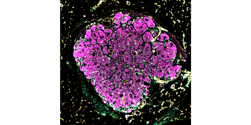
Image credit: Chelsea Fortin, Kelly Stevens and Sangeeta Bhatia
In a study published July 19 in Science Translational Medicine, researchers discovered that a “seed” of human liver and supporting cells “blossomed” to 50 times its original size in mice. The cell structure integrated with the mouse circulatory system, and began to perform a few liver functions. The experiments were led by UW Bioengineering Assistant Professor Kelly Stevens when she was a postdoc at MIT.
To build the seeds, the researchers assembled three different types of human cells, primary heptocytes, umbilical vein endothelial cells and fibroblasts, in a degradable hydrogel. They then implanted the tissue seed into a liver injury mouse model. The mice were missing an amino acid metabolism gene that ultimately results in progressive liver failure. The researchers hypothesized that this model would facilitate the seeds’ growth by producing signaling factors that would prompt the human liver tissue to regenerate.
The researchers’ work offers a direction for studying organ development and a potential strategy for organ engineering. Their approach could lead to clinical solutions for organ disease and failure. There is a particular need for liver transplant tissue, so such “seeds” of engineered tissue could be used as an alternative to whole organ transplantation.
The work was featured in:
US News and World Report