Joint Professor of Radiology and Bioengineering
kinahan@uw.edu
Phone: (206) 543-0236
Office: Portage Bay Building (PBB) Room 222
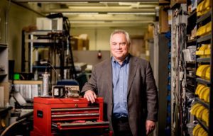
Paul Kinahan
Several of our developments are now included in hospital imaging systems and research systems worldwide. We collaborate closely with industry, physicians, and other researchers on pushing the envelope for direct improvement of patient welfare.
Image reconstruction and analysis algorithms
Quantitative imaging for treatment planning and assesing response to therapy
Measuring and improving medical image quality.
B.A.Sc. Engineering Physics, University of British Columbia, Vancouver, BC 1985
M.A.Sc. Engineering Physics, University of British Columbia, Vancouver, BC 1988
Thesis “Three Dimensional Image Reconstruction”. Advisor Joel Rogers, PhD
Ph.D. Bioengineering, University of Pennsylvania, Philadelphia, PA 1994
Thesis “Image Reconstruction Algorithms for Volume Imaging PET Scanners” Advisor: Joel Karp, PhD
BC Science Council G.R.E.A.T. Graduate Fellowship
Student Fellowship Award, The Society of Nuclear Medicine
Graduate Scholarship Award, IEEE Nuclear and Plasma Sciences Society
Visiting Professorship: Herchel Smith Laboratory, University of Cambridge
IEEE-NPSS Young Investigator Medical Imaging Science Career Award
Visiting Professorship: National Institute of Radiological Sciences. Chiba, Japan
Fellow, Institute of Electrical and Electronics Engineers
Permanent Membership. IEEE Nuclear and Plasma Sciences Society
Edward J. Hoffman Award, The Society of Nuclear Medicine
Visiting Professorship. Department of Bio-Convergence. Korea University. Seoul, Korea
Visiting Professorship. Department of Physics, University of Sao Paolo, Brazil
Distinguished Lecture, Emory University Department of Radiology
Fellow, American Association of Physicists in Medicine
BIOE 599 – Fundamentals of Biomedical Imaging: X-ray and Nuclear Biomedical Imaging.
Explores core principles of biomedical imaging with a focus on x-ray and nuclear imaging. Fundamental concepts common to all modalities are reviewed: Multi-dimensional Fourier transforms, the imaging equation, the inverse problem, image SNR, and contrast agents. Lectures will emphasize a systems approach that is reinforced though computational mini projects using Matlab or equivalent.
Deep-learning derived features for lung nodule classification with limited datasets. P Thammasorn, W Wu, LA Pierce, SN Pipavath, PD Lampe, AM Houghton, PE Kinahan. Medical Imaging 2018: Computer-Aided Diagnosis, SPIE 2018.
A phantom design for assessment of detectability in PET imaging. SD Wollenweber, AM Alessio, PE Kinahan. Medical physics 43 (9), 5051-5062 2016.
Radiomics: images are more than pictures, they are data. RJ Gillies, PE Kinahan, H Hricak. Radiology 278 (2), 563-577. 2015.
In the News
Three UW Bioengineering Professors elected to join the Washington State Academy of Sciences
2024-08-16T07:08:04-07:00August 16th, 2024|
Paul Kinahan and Cross-Organizational Team Awarded ARPA-H Funding
2023-11-20T11:45:07-08:00November 20th, 2023|
Asst. Professor Kelly Stevens and Prof. Paul Kinahan named AIMBE Fellows
2022-02-18T13:23:16-08:00February 18th, 2022|
UW Bioengineers pivot to develop coronavirus solutions
2022-08-01T14:43:01-07:00July 9th, 2020|


