

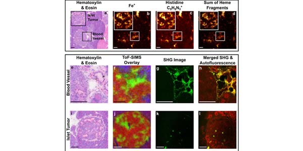
Image: H&E, ToF-SIMS, and SHG images of vasculature and blood lakes. View full image description.
Analysis of the Myc-induced pancreatic B cell islet tumor microenvironment using imaging ToF-SIMS
Blake M. Bluestein, Fionnuala Morrish, Daniel J. Graham, Li Huang, David Hockenbery, and Lara J. Gamble
Biointerphases. Volume 13, 06D402 (2018).
Abstract
Solid tumors are a structurally complex system, composed of many different cell types. The tumor microenvironment includes nonmalignant cell types that participate in complex interactions with tumor cells. The cross talk between tumor and normal cells is implicated in regulating cell growth, metastatic potential, and chemotherapeutic drug resistance. A new approach is required to interrogate and quantitatively characterize cell to cell interactions in this complex environment. Here, the authors have applied time-of-flight secondary ion mass spectrometry (ToF-SIMS) to analyze Myc-induced pancreatic B cell islet tumors. The high mass resolution and micron spatial resolution of ToF-SIMS allows detection of metabolic intermediates such as lipids and amino acids. Employing multivariate analysis, specifically, principal component analysis, the authors show that it is possible to chemically distinguish cancerous islets from normal tissue, in addition to intratumor heterogeneity. These heterogeneities can then be imaged and investigated using another modality such as sum harmonic generation microscopy. Using these techniques with a specialized mouse model, the authors found significant metabolic changes occurring within B cell tumors and the surrounding tissues. Specific alterations of the lipid, amino acid, and nucleotide metabolism were observed, demonstrating that ToF-SIMS can be utilized to identify large-scale changes that occur in the tumor microenvironment and could thereby increase the understanding of tumor progression and the tumor microenvironment.