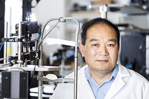
Professor of Bioengineering and Ophthalmology
George And Martina Kren Endowed Chair In Ophthalmology Research
wangrk@uw.edu
Phone: (206) 616-5025
Office: Foege N410E
Ruikang (Ricky) Wang
Functional optical imaging using coherence gating (OCT) and confocal gating techniques
Photoacoustic imaging
Laser Doppler, speckle and intrinsic optical signal imaging
Optical biopsy and functional imaging in tissue engineering
Light propagation in biological tissue
M.Sc., Tianjin University, 1990
B.Eng., Tianjin University, 1988
Fellow of Optical Society of America (OSA)
Fellow of International Society of Optics and Photonics (SPIE)
Recipient of Research to Prevent Blindness Innovative Research Award
Recipient of OHSU Technology Innovation Award
Editor of Journal of Biomedical Optics Letters
Associate Editor for Biomedical Optics Express, Quantitative Imaging in Medicine and Surgery
BIOEN 599: Biomedical Imaging
BIOEN 498/599: Optical coherence tomography
JR de Oliveira Dias, QQ Zhang, JMB Garcia, F Zheng, EH Motulsky, L Roisman, AR Miller, CL Chen, S Kubach, L de Sisternes, MK Durbin, RK Wang, G Gregori, PJ Rosenfeld. “Prevalence and Natural History of Subclinical Neovascularization in Age-Related Macular Degeneration Imaged with Swept Source OCT Angiography.” Ophthalmology, (In Press, 2017).
A.H. Kashani, C.-L. Chen, J.K. Gahm, F. Zheng, G.M. Richter, P.J. Rosenfeld, Y. Shi, R.K. Wang, “Optical coherence tomography angiography: A comprehensive review of current methods and clinical applications”, Progress in Retinal and Eye Research (In press, 2017), doi: 10.1016/j.preteyeres.2017.07.002.
RK Wang, QQ Zhang, YD Li, and SZ Song. “Optical coherence tomography angiography based capillary velocimetry.” J Biomed Opt. 22(6), 066008 (Jun 15, 2017).
QQ Zhang, CL Chen, ZD Chu, F Zheng, AR Miller, L Roisman, JR De Oliveira Dias, Z Yehoshua, KB. Schaal, W Feuer, G Gregori, S Kubach, L An, PF Stetson, MK Durbin, PJ Rosenfeld, RK Wang. “Automated quantitation of choroidal neovascularization: a comparison study between spectral domain and swept source OCT angiograms.” Investigative Ophthalmology & Visual Science, 58 (3):1506– 1513 (2017). DOI:10.1167/iovs.16-20977.
CL Chen, KD Bojikian, JC Wen, QQ Zhang, C Xin, RC Mudumbai, MA Johnstone, PP Chen, RK Wang “Peripapillary Retinal Nerve Fiber Layer Vascular Microcirculation in Glaucomatous Eyes with Single Hemifield Visual Field Loss.” JAMA Ophthalmology. 135(5):461-468 (2017) doi:10.1001/jamaophthalmol.2017.0261.
J.J. Xu, S.Z. Song, W. Wei and R.K. Wang, “Wide field and highly sensitive angiography based on optical coherence tomography with akinetic swept source.” Biomedical Optics Express, 8 (1), 420-435 (2017). https://doi.org/10.1364/BOE.8.000420.
CL Chen, and RK Wang. “Optical Coherence Tomography Based Angiography” Biomedical Optics Express, 8(2), 1056 – 1082 (2017).
L. Ambrozinski*, S. Song*, S. J. Yoon*, I. Pelivanov, D. Li, L. Gao, T.T. Shen, R.K. Wang and M. O’Donnell. “Acoustic micro-tapping for non-contact 4D imaging of tissue elasticity. Sci. Rep. 6, 38967; doi: 10.1038/srep38967 (2016).
WJ Choi, YD Li, W Qin and RK Wang. “Cerebral capillary velocimetry based on temporal OCT speckle contrast”. Biomedical Optics Express, 7(12), 4859-4873 (2016). https://doi.org/10.1364/BOE.7.004859
S.Z. Song, J.J. Xu and R.K. Wang, “Long-range and wide field of view optical coherence tomography for in vivo 3D imaging of large volume object based on akinetic programmable swept source.” Biomedical Optics Express, 7 (11): 4734-4748 (2016). https://doi.org/10.1364/BOE.7.004734
Y. Nishijima, Y. Akamatsu, S.Y. Yang, C.C. Lee, U. Baran, S. Song, R.K. Wang, T. Tominaga, J.L. Liu, “Impaired collateral flow compensation during chronic cerebral hypoperfusion in the type-II-diabetic mice”, Stroke 47(12):3014-3021(2016). DOI: 10.1161/STROKEAHA.116.014882
Q.Q. Zhang, A.Q. Zhang, C. Lee, A. Lee, K.A. Rezaei, A. Miller, F. Zheng, L. Roisman, G. Gregori, M.K. Durbin, L. An, P. F. Stetson, P. J. Rosenfeld and R.K. Wang. “Projection artifact removal enables accurate presentation and monitoring of choroidal neovascularization imaged by optical coherence tomography angiography”, Ophthalmology Retina, 1:124-136 (2017).
C.L. Chen, A.Q. Zhang, Q.Q. Zhang, D. Gupta, J.C. Wen, C. Xin, R.C. Mudumbai, K.D. Bojikian, M.A. Johnstone, P.P. Chen, R.K. Wang. “Peripapillary Retinal Nerve Fiber Layer Vascular Microcirculation in Glaucoma Using Optical Coherence Tomography–Based Microangiography”, Investigative Ophthalmology & Visual Science, 57, OCT475-OCT485 (2016). doi:10.1167/iovs.15-18909.
L. Warren*, S. Ramamoorthy*, N. Ciganovic*, Y. Zhang, T. Wilson, T. Petrie, R.K. Wang, S.L. Jacques, T. Reichenbach, A.L.Nuttall, A. Fridberger. “Minimal basilar membrane motion in low-frequency hearing.” Proc Natl Acad Sci USA. 113(30):E4304-10. (2016)
ZD Chu, J. Lin, C. Gao, C Xin, Q Zhang, C-L Chen, L. Roisman, G Gregori, PJ Rosenfeld, and R.K. Wang. “Quantitative assessment of the retinal microvasculature using OCT angiography”, Journal of Biomedical Optics, 21(6), 066008 (2016), doi: 10.1117/1.JBO.21.6.066008.
S.Z. Song, W. Wei, B.Y. Hsieh, I. Pelivanov, T.T. Shen, M. O’Donnell and R.K. Wang. “Strategies to improve phase-stability of ultrafast swept source optical coherence tomography for single shot imaging of transient mechanical waves at 16 kHz frame rate”. Applied Physics Letters. 108, 191104 (2016); doi: 10.1063/1.4949469.
C. Xin, R.K. Wang, S.Z. Song, T.T. Shen, J. Wen, E. Martin, Y. Jiang, S. Padilla, M. Johnstone. “Aqueous outflow regulation: Optical coherence tomography implicates pressure-dependent tissue motion.” Experimental Eye Research, 158 (5):171-186. (2017). http://dx.doi.org/10.1016/j.exer.2016.06.007
Q.Q. Zhang, C.S. Lee, J. Chao, C.L. Chen, T. Zhang, U. Sharma, A.Q. Zhang, J. Liu, K. Rezaei, K.L. Pepple, R. Munsen, J. Kinyoun, M. Johnstone, R.N. Van Gelder, and R.K. Wang. “Wide-field optical coherence tomography based microangiography for retinal imaging”, Scientific Reports, 6, Article number: 22017 (2016). doi:10.1038/srep22017.
L. Roisman, Q.Q. Zhang, R.K. Wang, G. Gregori, A. Zhang, C.L. Chen, M.K. Durbin, L. An, P. F. Stetson, G. Robbins, A. Miller, F. Zheng, and P. J. Rosenfeld “Optical Coherence Tomography Angiography of Asymptomatic Neovascularization in Intermediate Age-Related Macular Degeneration”. Ophthalmology, 123(6):1309 – 1319 (2016). doi:10.1016/j.ophtha.2016.01.044
U. Baran, W Qin, X. Qi, G Kalkan, and R.K. Wang. “OCT-based label free in vivo lymphangiography within human skin”, Scientific Reports, 6, 21122 (2016).
In the News
Imaging pioneer Ruikang Wang to deliver Inaugural Fujimoto Lecture
2025-03-31T11:31:26-07:00March 31st, 2025|
Ruikang Wang is recognized for transforming eye care with 3D blood flow imaging
2025-01-23T10:33:31-08:00January 23rd, 2025|
Three UW Bioengineering Professors elected to join the Washington State Academy of Sciences
2024-08-16T07:08:04-07:00August 16th, 2024|
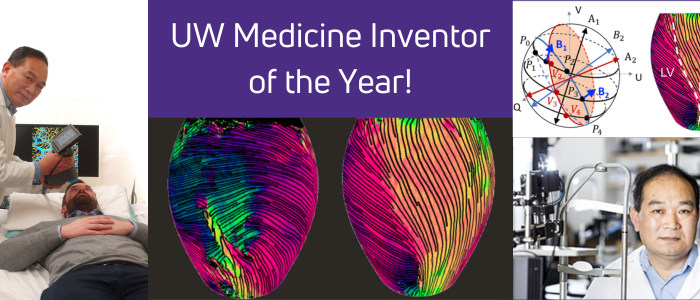
Ruikang Wang is 2023 UW Medicine Inventor of the Year
2023-10-17T05:51:34-07:00July 13th, 2023|
Ruikang Wang named editor of Biomedical Optics Express
2022-01-07T11:53:01-08:00January 5th, 2022|
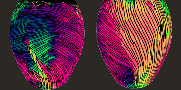
Imaging method captures deep layers of collagen in 3D
2021-12-17T11:13:45-08:00December 16th, 2021|


