
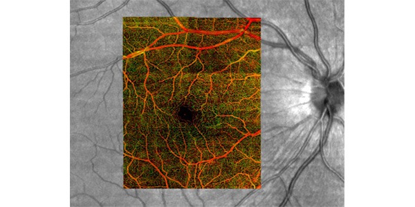
Photo: MICROCIRCULATION IN ACTION UW Bioengineering’s new non-invasive 3-D imaging technique (overlay, in color) reveals a multitude of tiny capillaries pumping blood to a human retina. Camera methods currently used in the clinic (black and white image of same retina) cannot capture such detail and carry risks.
UW Bioengineer Ruikang Wang’s non-invasive method for imaging vascular health holds promise for better diagnosis, monitoring and treatment of diseases.
The body relies on its network of vascular highways and byways t oshuttle blood to remote outposts, where it nourishes cells and sweeps away waste in order to manage inflammation, heal a wound or repair other damage. A new way of looking at blood vessels, pioneered by Ruikang Wang, professor of bioengineering, and his lab, is shedding light on vascular processes in the tiny world of the microcirculatory system.
The system has been used to map vascular networks and image blood movement in living tissues such as human eyes and skin, and mouse brains and kidneys. The highly sensitive imaging can also detect more subtle changes in mechanical properties of tissues than current methods, and may one day allow diseases to be imaged or detected at an earlier stage than what’s possible now, Wang says.
The team’s high resolution imaging technique, called optical microangiography, works on the same principle as ultrasound imaging, but rather than using sound waves to detect changes in structure, the new system uses light. The device shines near-infrared light on the area to be scanned, and then measures the light that bounces back in a number of ways. The team’s algorithms compile an ultra-fine 3-D map of the functioning blood vessel network and surrounding tissue. The resolution is so fine that it can separate signals that are backscattered from moving particles, such as blood cells in vessels, from non-moving particles in other tissue – no fluorescent contrast dye needed. The result is a real-time, dye-free and non-invasive imaging technique that tracks blood flow and speed in functional vessels down to the capillary level – as small as five micrometers in diameter.
The technology can reach as deep as three millimeters into tissue – deep enough to scan the eye’s retina, or see into the layer of skin or tissue where inflammation occurs.
Wang’s team is studying a number of uses for the new technique. Their system has helped explain the causes of tissue death in certain cosmetic surgery procedures, and how removing outer layers of dead skin cells activates reserve blood vessels to speed drug delivery through topical creams. In one study, they measured how quickly blood rushed back into a fingertip after first applying pressure to stop the flow of blood and then releasing it – a potentially useful measure for diagnosing or evaluating diabetes, pressure ulcers and stroke.
Window into eye health
The team’s research may make it easier to diagnose and treat eye diseases, especially those involving blood vessels in the retina, in which the vessels become swollen, leaky or grow abnormally.
Age-related macular degeneration, or AMD, is the leading cause of vision loss in Americans over age 60. AMD gradually destroys the macula, a cluster of light-sensitive cells in the central part of the retina. It comes in two forms – dry and the more advanced wet – and is named according to how the damage occurs. In dry AMD, the macula breaks down and photoreceptors die, causing cloudy central vision or a central area of vision loss. In the wet form, new blood vessels behind the retina start to grow under the macula. These abnormal vessels tend to be fragile, and can easily leak blood or fluid, damaging the macula, and also causing cloudy or blurry central vision, usually worse than in the dry form.
“It is not known how vasculature function is involved in AMD,” Wang says. “The vascular involvement in this disease is clear, but we are handicapped by our ability to visualize and localize the tiny capillaries within the retina. We’re hoping that our imaging system will shed some light on the mechanism of AMD development and progression.”
People with diabetes need to carefully monitor their eye health, as prolonged high blood sugar levels can eventually damage the fine vascular network in the retina. Diabetic retinopathy tops the list of leading causes of blindness among working Americans ages 18-65. Damage to the capillaries can cause leaking and decrease the blood supply to the retina, starving the tissue of oxygen and nutrients. The weakened capillaries form bell-shaped protrusions on the capillary walls called microaneurysms, the earliest clinical sign of diabetic retinopathy. Currently the only way to monitor the abnormal capillary changes is to inject a fluorescent dye into the patient’s veins, wait for it to travel through the bloodstream to the eye, and take rapidphotographs just as the dye passes through the retinal capillaries. The timing can be difficult, Wang notes. “Our technique doesn’t need a contrast agent,” Wang says, and it avoids the cost and potential complications of using such dyes. “We can visualize very precisely the formation of microaneurysms, and we can measure the diameter of the capillaries and monitor changes over time, as needed. We have the potential to catch problems earlier than previously possible.”
Wang collaborates with a team from the UW Department of Ophthalmology, including Russ Van Gelder, chair and professor, Jennifer Chao, assistant professor, James Kinyoun, professor, and Murray Johnstone, clinical professor, to study the benefits of the imaging technique. The clinicians are currently testing the system with patients and comparing its resolution to current methods. Wang and his team’s initial results show that the new technique provides a more detailed image than current ophthalmic cameras (see image).
“Dr. Wang’s new technology shows a lot of promise for being able to identify disease at avery early stage and to monitor it on a regular and frequent basis – something we’ve never had available to us before,” Kinyoun says. “The way we’ve been following patients with vessel problems, such as diabetic retinopathy, involves injection of a dye into someone’s blood stream, and there’s always a small risk of serious complications due to that dye.Now we have a non-invasive procedure that can be done with much less risk that can give us even more information than what we’ve hadin the past. So I think it will enable us to pick up disease much earlier, to understand it better and to find better treatments.”
Wang’s lab partnered with optics company Carl Zeiss on a prototype device for ophthalmologists. “We don’t see any hurdles to moving this into the clinical system because this is based on optical coherence tomography (OCT) technology that is used in every clinic already,” Wang says. “This is really an add-on technique to current OCT systems. The biggest difference is the mathematics – the algorithm software.”
Potential to image brain, heart, lung, digestive disease
Wang believes the new technology could benefit a range of areas affected by blood flow and to help in early diagnosis, monitoring of a condition and treatment of diseases. The team is studying the benefit of using the system to monitor the effects of stroke and traumatic brain injury. Experiments in live mice detail the blood flow dynamics and ability of new vessels to form in the brain.
The technique could be easily added to an existing catheter to look at cardiovascular function in heart disease, Wang says. Currently doctors thread a catheter through a vessel to look at plaques in the arteries. “This is a very difficult task,” Wang says. “One of most important factors to look at with vulnerable plaque is to look at new vessels forming beneath the plaque.” If the plaque is fragile, it could break off the artery wall and be swept into the bloodstream. The improved imaging could guide clinicians on how to best treat the plaque.
Wang envisions it could also be attached to an endoscope to evaluate the digestive system and perhaps look at or help treat stomach cancer or colon cancer. It may one day aid in laparoscopic surgery or be added to a bronchoscope to better image airways and lungs.