
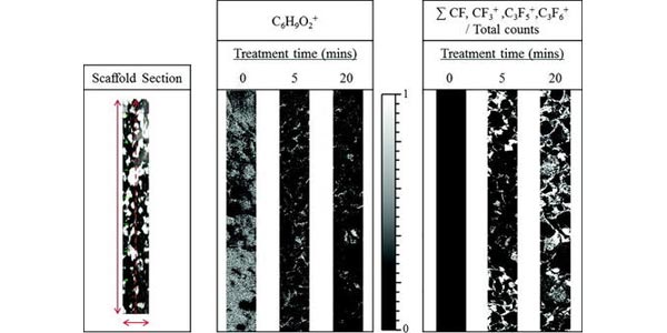
Image in the first column is a representative photomicrograph of an unmodified PCL scaffold, the direction of analysis for the corresponding secondary ion images is detailed by the red dashed line (500?×?4000??m). The second and third columns are ToF-SIMS image area scans (500?×?4000??m) from cross-sections of C3F8 plasma treated (0, 5, and 20?min) PCL scaffolds following the C6H9O2+ ion (PCL substrate) and a sum of FC ions from the plasma deposition film.
Taylor MJ, Aitchison H, Hawker MJ, Mann MN, Fisher ER, Graham DJ, Gamble LJ.
Biointerphases. 2018 Mar 30;13(3):03B415.
Abstract
Biopolymers are used extensively in the manufacture of porous scaffolds for a variety of biological applications. The surfaces of these scaffolds are often modified to encourage specific interactions such as surface modification of scaffolds to prevent fouling or to promote a cell supportive environment for tissue engineering implants. However, few techniques can effectively characterize the uniformity of surface modifications in a porous scaffold. By filling the scaffold pores through polymer embedding, followed by analysis with imaging time-of-flight secondary ion mass spectrometry (ToF-SIMS), the distribution and composition of surface chemical species though complex porous scaffolds can be characterized. This method is demonstrated on poly(caprolactone) scaffolds modified with a low-fouling plasma-deposited coating from octafluoropropane via plasma enhanced chemical vapor deposition. A gradient distribution of CF+/CF3+ is observed for scaffolds plasma treated for 5?min, whereas a 20?min treatment results in more uniform distribution of the surface modification throughout the entire scaffold. The authors expect this approach to be widely applicable for ToF-SIMS analysis of scaffolds modified by multiple plasma processing techniques as well as alternative surface modification approaches.