
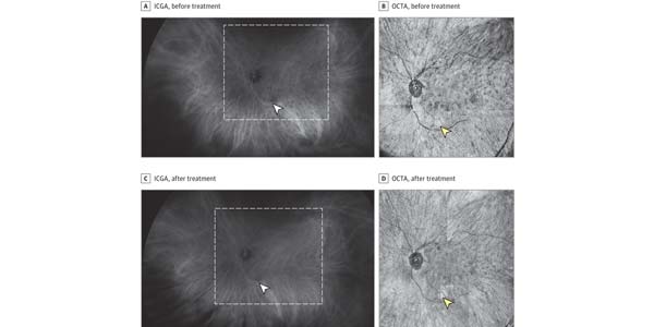
Image: Identification of Choroidal Flow Voids Corresponding to Indocyanine Green Angiography (ICGA) Lesions via Swept-Source Optical Coherence Tomography Angiography (SS-OCTA)
Kathryn L. Pepple, MD, PhD; Zhongdi Chu, MS; Jessica Weinstein, MD; Marion R. Munk, MD, PhD; Russell N. Van Gelder, MD, PhD; Ruikang K. Wang, PhD1,2
JAMA Ophthalmology, 2018;136(11):1288-1292.
Question
Can noninvasive swept-source optical coherence tomography angiography detect deep choroidal inflammatory lesions?
Findings
In this case series of 3 patients with birdshot chorioretinopathy, widefield imaging with swept-source optical coherence tomography angiography identified areas of abnormal flow signal, termed flow voids, in the choroid in a pattern similar to indocyanine green angiography. Furthermore, serial en face images obtained at different choroidal depths suggest acute lesions localize to Haller layer, while chronic lesions may involve the entire choroid.
Meaning
En face analysis of widefield swept-source optical coherence tomography angiography imaging shows promise as a noninvasive method for detecting and monitoring choroidal inflammatory diseases such as birdshot chorioretinopathy.