UW Bioengineering
Fast Facts
News and Events

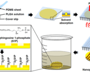
MAB alumnus raises $5 million to build autonomous surgical robotics
Georgia Witchel and the team at Louiza Labs has raised $5million to develop the technology underlying simulated FDA trials and autonomous surgical robots.
Events

Three UW Bioengineering Professors elected to join the Washington State Academy of Sciences
Three UW BioE faculty members have been selected to join the Washington State Academy of Sciences (WSAS): Valerie Daggett, Paul Kinahan and Ruikang Wang. T
HHMI Gilliam Fellowship highlights the enduring power of mentorship
Sydney Floryanzia, Ph.D. student, and Elizabeth Nance, an associate professor, have been recognized as a 2024 HHMI Gilliam Fellowship student-advisor pair.

Kelly Stevens co-leads new NIH-funded center to reduce disparities in biomaterials research
The National Institutes of Health is supporting a new center to advance biomaterials research and connect researchers with a grant of $10.5 million.
News & Events


Featured Publications
Human embryonic stem cell–derived cardiomyocytes restore function in infarcted hearts of non-human primates
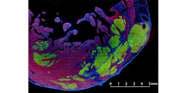
Charles Murry and colleagues demonstrate that remuscularization of the infarcted macaque heart with human myocardium provides durable improvement in left ventricular function.
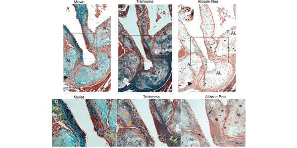
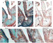
Increased Calcific Aortic Valve Disease in response to a diabetogenic, procalcific diet in the LDLr-/-ApoB100/100 mouse model
The Scatena and Giachelli labs developed an animal model that mimicked the structural and functional features of CAVD in people with T2DM, by testing a diabetogenic, procalcific diet and its effect on the incidence and severity of CAVD and AS in the, LDLr-/-ApoB100/100 mouse model.
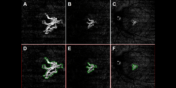
Comparison of Neovascular Lesion Area Measurements From Different Swept-Source OCT Angiographic Scan Patterns in Age-Related Macular Degeneration
The researchers compared area measurements for the same neovascular lesions imaged using swept source optical coherence tomography angiography (SS-OCTA) and enlarging scan patterns. The similarity in lesion area measurements across different scan patterns suggests that SS-OCTA imaging can be used to follow quantitatively the enlargement of choroidal neovascularization as the disease progresses.