

Single-cell profiling of the developing mouse brain and spinal cord with split-pool barcoding
Researchers from Georg Seelig’s (Electrical Engineering, adjunct BioE) and Suzie Pun/Drew Sellers’ labs, and the Allen Institute for Brain Science, have developed a new single-cell RNA sequencing method that can reliably track gene activity in a tissue sample to the individual cell level.
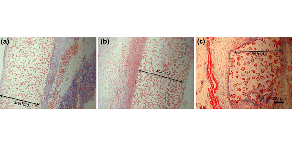
Precision-porous templated scaffolds of varying pore size drive dendritic cell activation
The Bryers labs presents the effects of varying pore size of poly (2-hydroxyethyl methacrylate) (pHEMA) and poly(dimethylsiloxane) (PDMS, silicone) scaffolds on the maturation and in vivo enrichment of dendritic cells.
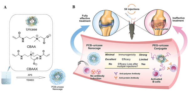
Zwitterionic Nanocages Overcome the Efficacy Loss of Biologic Drugs
Researchers from Shaoyi Jiang's (BioE adjunct) lab demonstrate a zwitterionic polycarboxybetaine nanocage that can physically encase proteins while keeping their structure intact. This approach addresses the problem posed by efficacy loss of biotherapeutics due to undesirable immune response.
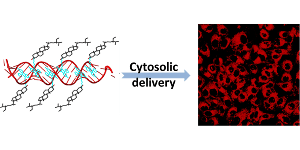
Noncovalent tagging of siRNA with steroids for transmembrane delivery
The Gao lab reports on the development of small, bifunctional chemical tags capable of transporting siRNA directly into the cytosol. The bifunctional tags consist of a siRNA-binding moiety that interacts with siRNA non-covalently, and a steroid domain that readily fuses with the mammalian cell membrane.

Harnessing Sphingosine-1-Phosphate Signaling and Nanotopographical Cues
Deok-Ho Kim's lab demonstrates that biomimetic nanotopography and S1P can be combined to synergistically regulate the maturation and vascularization of engineered skeletal muscles.
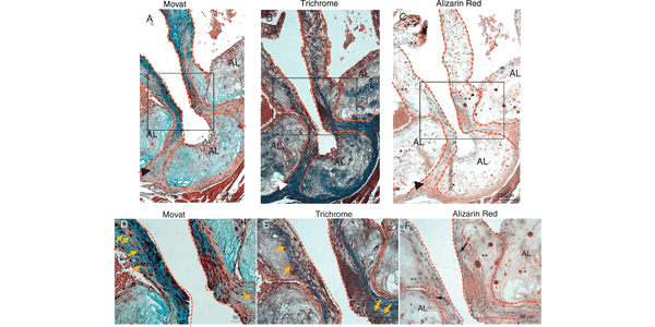
Increased Calcific Aortic Valve Disease in response to a diabetogenic, procalcific diet in the LDLr-/-ApoB100/100 mouse model
The Scatena and Giachelli labs developed an animal model that mimicked the structural and functional features of CAVD in people with T2DM, by testing a diabetogenic, procalcific diet and its effect on the incidence and severity of CAVD and AS in the, LDLr-/-ApoB100/100 mouse model.